Confocal Microscopy Image Gallery
Rat Brain Tissue Sections
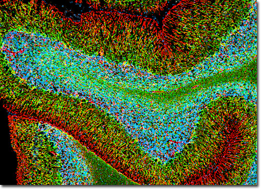
Cerebellum
|
The cerebellum is a highly organized brain structure, as demonstrated by the image of a rat brain coronal section presented above. The specimen was immunofluorescently labeled for astrocytes and neurons with rabbit anti-GFAP monoclonal antibodies and chicken anti-NF-H antibodies followed by goat anti-rabbit and anti-chicken secondary antibodies conjugated to Alexa Fluor 568 and Alexa Fluor 488, respectively. GFAP is strongly and specifically expressed by various astroglia, and NF-H is solely found in the neurofilaments of neuronal cells. Cell nuclei were visualized with the red-absorbing probe DRAQ5 (pseudocolored cyan). Images were recorded with a 10x objective using a zoom factor of 1.0 and sequential scanning with the 488-nanometer spectral line of an argon-ion laser, the 543-nanometer line from a green helium-neon laser, and the 633-nanometer line of a red helium-neon laser. During the processing stage, individual image channels were pseudocolored with RGB values corresponding to each of the fluorophore emission spectral profiles unless otherwise noted above. View a larger version of this digital image. |
 |
 |
 |
 |
 |
 |
 |