 |
 |
 |
|
||||||||||||||||||||||||
 | ||||||||||||||||||||||||
 | ||||||||||||||||||||||||
 | ||||||||||||||||||||||||
Confocal Microscopy Image Gallery
Mouse Hemangioendothelioma Endothelial Cells (EOMA Line)
A hemangioendothelioma is any of a broad group of tumors that arise from the endothelium of a blood vessel. The epithelial EOMA cell line was initiated from mixed hemangioendothelioma tissue excised from an adult mouse (Mus musculus).

EOMA cells express vascular addressin (an endothelial cell adhesion molecule) and surface receptors for acetylated low-density lipoprotein. An array of cellular products are synthesized by the cells, including Cathepsin L, endostatin, interleukin-6, angiotensin-converting enzyme (ACE), and thrombospondin. Studies with syngeneic mice indicate that the EOMA line is tumorigenic.
Hemangioendotheliomas often have the appearance of red or blue nodules and are frequently malignant. Their behavior is typically classified somewhere between that of a benign hemangioma and a malignant angiosarcoma. The neoplasms are not well circumscribed and several lesions may be present. Around 10 percent of diagnosed cases are linked to lymphedema, Maffucci's syndrome, varicose veins or other conditions. Hemangioendotheliomas do not often occur in children and affect adult men and women at similar rates. Treatment and prognosis for the tumors vary based on a number of factors, such as the specific type of hemangioendothelioma present, location of the growth, and the general health of the patient.
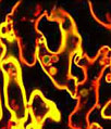
A log phase culture of mouse endothelial cells was immunofluorescently labeled with primary anti-tubulin mouse monoclonal antibodies followed by goat anti-mouse secondary antibodies conjugated to Alexa Fluor 555 (pseudocolored blue). The cells were simultaneously stained for DNA with TO-PRO-3, a carbocyanine monomer with long-wavelength red fluorescence. Images were recorded with a 60x oil immersion objective using a zoom factor of 2.5 and sequential scanning with the 543-nanometer line from a green helium-neon laser and the 633-nanometer line of a red helium-neon laser. During the processing stage, individual image channels were pseudocolored with RGB values corresponding to each of the fluorophore emission spectral profiles unless otherwise noted above.
Additional Confocal Images of Mouse Endothelial (EOMA) Cells
Targeting the Microtubule Network in Mouse Endothelial Cells with Immunofluorescence - The EOMA endothelial cell shown in this confocal image was resident in a culture that was fixed, permeabilized, blocked, and then treated with mouse primary antibodies that target alpha-tubulin, a major component of the microtubule network. The primaries were labeled with goat anti-mouse secondary antibodies conjugated to Alexa Fluor 555 in a solution containing TO-PRO-3 to simultaneously stain DNA.
Contributing Authors
Nathan S. Claxton, Shannon H. Neaves, and Michael W. Davidson - National High Magnetic Field Laboratory, 1800 East Paul Dirac Dr., The Florida State University, Tallahassee, Florida, 32310.