 |
 |
 |
|
||||||||||||||||||||||||
 | ||||||||||||||||||||||||
 | ||||||||||||||||||||||||
 | ||||||||||||||||||||||||
Confocal Microscopy Image Gallery
Mink Uterus Endometrium Epithelial Cells (GMMe Line)
The GMMe cell line was initiated via the stable transfection of tissue excised from the endometrial layer of an adult minks uterus. The plasmid vector utilized for the transfection encoded simian virus 40 (SV40) large tumor antigen (T antigen).

The cells were then cotransfected with another plasmid vector containing the gene for neomycin resistance and selected in a medium containing the antibiotic G418. GMMe cells exhibit epithelial morphology and have been used in co-culture with mink embryos in obligate diapause to improve their rate of survival in vitro, although a mink stromal line, GMMs, was used for the same purpose with more success. Testing indicates that the epithelial cells are positive for cytokeratin, vimentin, and alkaline phosphatase, but negative for desmin.
A central component of the female mammalian reproductive system, the uterus is a pear-shaped muscular organ that typically lies in the pelvis. During pregnancy, the uterus, also known as the womb, expands greatly, filling a much larger space in the body as the fetus it holds grows and develops. The wall of the uterus consists of several layers of tissue, the innermost of which is the endometrium, a glandular mucus membrane. In addition to glands, the endometrium contains blood vessels and lymphatic spaces. During pregnancy, the blood vessels of the endometrium are connected to the embryo so that a placenta is formed. When females of reproductive age are not pregnant, parts of the endometrium are shed and then regenerated as part of the menstrual cycle.
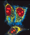
The Golgi apparatus was targeted in the culture of mink endometrium epithelial cells appearing in the digital image above by treating the cells with rabbit (anti-giantin) primary antibodies, followed by secondary antibodies conjugated to Alexa Fluor 568 (pseudocolored cyan). The culture was also stained for mitochondria with MitoTracker Deep Red 633, and for the actin cytoskeleton with Alexa Fluor 488 conjugated to phalloidin. Images were recorded with a 60x oil immersion objective using a zoom factor of 2.0 and sequential scanning with the 488-nanometer spectral line of an argon-ion laser, the 543-nanometer line from a green helium-neon laser, and the 633-nanometer line of a red helium-neon laser. During the processing stage, individual image channels were pseudocolored with RGB values corresponding to each of the fluorophore emission spectral profiles unless otherwise noted above.
Additional Confocal Images of Mink Uterus Endometrium Epithelial (GMMe) Cells
Distribution of Histones and Peroxisomes in Mink Endometrium Epithelial Cells - A log phase culture of GMMe cells was treated with a cocktail of mouse anti-histones (pan) and rabbit anti-PMP 70 (peroxisomal membrane protein) primary antibodies, followed by goat anti-mouse and anti-rabbit secondary antibodies conjugated to Texas Red and Alexa Fluor 488, respectively. The procedure enabled the visualization of the nuclear histone proteins (red emission) and cytoplasmic peroxisomes (green emission) present in the cells.
Contributing Authors
Nathan S. Claxton, Shannon H. Neaves, and Michael W. Davidson - National High Magnetic Field Laboratory, 1800 East Paul Dirac Dr., The Florida State University, Tallahassee, Florida, 32310.