 |
 |
 |
|
||||||||||||||||||||||||
 | ||||||||||||||||||||||||
 | ||||||||||||||||||||||||
 | ||||||||||||||||||||||||
Confocal Microscopy Image Gallery
Human Embryonic Kidney Epithelial Cells (HEK 293 Line)
Human embryonic kidney (HEK) cells have been used in scientific research for many years regardless of the controversy that surrounds them and other cell lines derived from human embryos. Many scientists that have exploited HEK cells have been exploring ways to utilize them to grow new kidneys in adults that have been diagnosed with renal failure, an undertaking that has been remarkably successful.

Since the late 1970s, HEK cells have also been commonly used in studies of adenoviruses, a collection of DNA-containing viruses that cause upper respiratory tract infections and pinkeye in humans. This additional application of the HEK line was made possible by the transformation of the cells with DNA from human adenovirus type 5 in 1977 by a group of scientists led by Frank Graham at McMaster University. To accomplish this feat, the group used a technique involving calcium phosphate precipitation. The result was a new cell line, generally referred to as HEK 293.
HEK 293 cells exhibit epithelial morphology and are well suited for cultivating and assaying human adenoviruses that had been difficult to produce and maintain in laboratory settings before the development of the cell line. Although some early reports suggested that the cells contained adenovirus 5 DNA from both the right and left ends of the viral genome, it is now widely accepted that only left end sequences are present. HEK 293 cells do not adhere to substrates when left at room temperature for any length of time. When kept at a temperature of 37 degrees Celsius, however, over a several day period, any loose cells in a flask or Petri dish become adherent. The cells express an unusual cell surface receptor for the adhesive protein vitronectin composed of the integrin beta-1 subunit and the vitronectin receptor alpha-v subunit, and studies indicate that they are tumorigenic in nude mice.
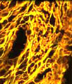
The distribution of filamentous actin and mitochondria in a culture of human embryonic kidney cells (shown above) was visualized with BODIPY FL conjugated to phallacidin and MitoTracker Red CMXRos (pseudocolored yellow). Cell nuclei were counterstained with TO-PRO-3, a carbocyanine monomer with long-wavelength red fluorescence. Images were recorded with a 60x oil immersion objective using a zoom factor of 2.5 and sequential scanning with the 488-nanometer spectral line of an argon-ion laser, the 543-nanometer line from a green helium-neon laser, and the 633-nanometer line of a red helium-neon laser. During the processing stage, individual image channels were pseudocolored with RGB values corresponding to each of the fluorophore emission spectral profiles unless otherwise noted above.
Contributing Authors
Nathan S. Claxton, Shannon H. Neaves, and Michael W. Davidson - National High Magnetic Field Laboratory, 1800 East Paul Dirac Dr., The Florida State University, Tallahassee, Florida, 32310.