 |
 |
 |
|
||||||||||||||||||||||||
 | ||||||||||||||||||||||||
 | ||||||||||||||||||||||||
 | ||||||||||||||||||||||||
Confocal Microscopy Image Gallery
Human Cervical Adenocarcinoma Cells (HeLa Line)
The HeLa cell line, which was initiated from a cervical adenocarcinoma, was the first successful immortal human cell line. Soon after the establishment of the epithelial line in the early 1950s, it was utilized in the development and testing of the polio vaccine.

HeLa cells are positive for the protein keratin and lysophosphatidylcholine (lyso-PC), which induces AP-1 activity and c-jun N-terminal kinase activity (JNK1) by a protein kinase C-independent pathway. According to genetic reports, HeLa cells contain human papilloma virus 18 (HPV-18) sequences and express p53 (tumor suppressor protein) at a low level. Levels of pRB (retinoblastoma suppressor) expression are normal. The proliferation of HeLa cells is remarkable, an entire generation of the cells being produced about once every day.
In addition to its role in making polio vaccinations a reality, the HeLa cell line has been used in research on such wide-ranging subjects as cancer, protein synthesis, radiation, and genetic control mechanisms. During the 1970s, however, HeLa cells were discovered to have been the cause of significant problems in a number of research laboratories. HeLa cells are so virulent that they were able to invade other cell cultures unbeknownst to the scientists utilizing them. As a result, many continuous human cell lines thought to have been developed after the HeLa line were actually simply HeLa cells. This momentous discovery can be largely credited to Walter Nelson-Rees, who was at that time co-director of the Cell Culture Laboratory at the Naval Biosciences Laboratory in Oakland, California. With his extensive karyotyping studies, Nelson-Rees revealed the astonishing extent of cross contamination of cell lines that had taken place, though many scientists, especially those whose research was discredited by the problem, were initially skeptical of his claim.
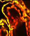
An adherent monolayer culture of human cervical adenocarcinoma cells was fixed, permeabilized, blocked, and then treated with mouse primary antibodies that target alpha-tubulin, a major component of the microtubule network. The primaries were labeled with goat anti-mouse secondary antibodies conjugated to Alexa Fluor 488 in a solution containing TO-PRO-3 to simultaneously stain DNA. Images were recorded with a 60x oil immersion objective using a zoom factor of 1.0 and sequential scanning with the 488-nanometer spectral line of an argon-ion laser and the 633-nanometer line of a red helium-neon laser. During the processing stage, individual image channels were pseudocolored with RGB values corresponding to each of the fluorophore emission spectral profiles.
Additional Confocal Images of Human Cervical Adenocarcinoma (HeLa) Cells
Tubulin, F-Actin, and DNA Distribution in HeLa Cells - The HeLa cell culture shown in this confocal image was labeled with mouse anti-alpha-tubulin primary antibodies followed by goat anti-mouse secondary antibodies conjugated to Alexa Fluor 488. In addition, the specimen was stained with Alexa Fluor 546 conjugated to phalloidin and TO-PRO-3, targeting the filamentous actin network and cell nuclei, respectively.
Visualizing the Proximity Between the Golgi Complex and the Nucleus in Carcinoma Cell Cultures - The Golgi apparatus was targeted in the culture of human cervical adenocarcinoma (HeLa) cells appearing in this section by treating the cells with rabbit (anti-giantin) primary antibodies, followed by secondary antibodies conjugated to Alexa Fluor 568. DNA in the cell nucleus was counterstained with the red-absorbing dye TO-PRO-3 (pseudocolored cyan).
EGFP-Peroxisome Subcellular Localization in Transfected Human Cervical Adenocarcinoma Cells - A culture of HeLa carcinoma cells was transfected with an EGFP-peroxisomal targeting signal 1 (PTS1) fusion protein and stained with MitoTracker Red CMXRos. These fluorescent probes respectively target the peroxisomes and intracellular microtubule network.
HeLa Cervical Carcinoma Cells Labeled with Alexa Fluor Dyes and TO-PRO-3 - Immunofluorescence with mouse anti-alpha-tubulin was employed to visualize details of the microtubule network in a log phase monolayer culture of HeLa adenocarcinoma cells. The secondary antibody (goat anti-mouse IgG) was conjugated to Alexa Fluor 488 and mixed with Alexa Fluor 546 conjugated to phalloidin to simultaneously image tubulin and the actin cytoskeleton. Nuclei were counterstained with TO-PRO-3, a carbocyanine monomer with long-wavelength red fluorescence.
Targeting Giantin in Cancer Cells with Immunofluorescence - The HeLa cell culture shown in this confocal image was fixed, permeabilized, washed, and blocked with 10-percent normal goat serum in phosphate-buffered saline prior to immunofluorescent labeling with rabbit primary antibodies to giantin, a protein resident in the Golgi complex of mammalian cells. The culture was subsequently stained with a mixture of secondary antibodies conjugated to Alexa Fluor 647. In addition, the filamentous actin network was probed with Alexa Fluor 488 conjugated to phalloidin, and DNA was labeled with the nucleic acid stain SYTOX Orange.
Contributing Authors
Nathan S. Claxton, Shannon H. Neaves, and Michael W. Davidson - National High Magnetic Field Laboratory, 1800 East Paul Dirac Dr., The Florida State University, Tallahassee, Florida, 32310.