 |
 |
 |
|
||||||||||||||||||||||||
 | ||||||||||||||||||||||||
 | ||||||||||||||||||||||||
 | ||||||||||||||||||||||||
Confocal Microscopy Image Gallery
Human Fetal Lung Fibroblast Cells (MRC-5 Line)
MRC-5 cells are a line of human lung fibroblast cells established in the 1960s by J. P. Jacobs. Studies have demonstrated that the cells are susceptible to several different viruses, including vesicular stomatitis (Indiana strain), poliovirus 1, and herpes simplex.

Cultured MRC-5 cells typically exhibit adherent growth and may double in population size as many as 46 times prior to senescence. The MRC-5 line is frequently used in laboratories for many different applications, such as in vitro cytotoxicity testing, the development of vaccines, and as a transfection host for investigations of viruses and viral diseases. MRC-5 cells are negative for reverse transcriptase.
In recent years, as the fight against terrorism has come to the forefront of American concerns, MRC-5 cells have experienced an increase in attention. This is because many modern smallpox vaccines are being generated by utilizing the MRC-5 line as opposed to the calf skin cell lines used to produce older versions of the vaccines. Smallpox vaccines have not been mandatory in the United States since the 1970s because the often fatal disease was already essentially eliminated from the country. Yet, since smallpox is highly contagious and few people are fully immune to it, a concerted effort is being made to ensure there are enough vaccine doses available in case the disease is ever reintroduced to the country via a biological weapon. Some Americans are, however, opposed to the utilization of MRC-5 cells for vaccine propagation, arguing against the scientific use of cells obtained from human fetal tissue.
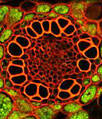
In a double immunofluorescence experiment, an adherent monolayer culture of MRC-5 cells (illustrated above) was fixed, permeabilized, blocked with 10 percent normal goat serum, and treated with a cocktail of mouse anti-vimentin and rabbit anti-PMP 70 (peroxisomal membrane protein) primary antibodies, followed by goat anti-mouse and anti-rabbit secondary antibodies (IgG) conjugated to Cy3 and Alexa Fluor 647 (pseudocolored blue), respectively. DNA in the cell nuclei was counterstained with SYTOX Green. Images were recorded with a 60x oil immersion objective using a zoom factor of 2.5 and sequential scanning with the 488-nanometer spectral line of an argon-ion laser, the 543-nanometer line from a green helium-neon laser, and the 633-nanometer line of a red helium-neon laser. During the processing stage, individual image channels were pseudocolored with RGB values corresponding to each of the fluorophore emission spectral profiles unless otherwise noted above.
Contributing Authors
Nathan S. Claxton, Shannon H. Neaves, and Michael W. Davidson - National High Magnetic Field Laboratory, 1800 East Paul Dirac Dr., The Florida State University, Tallahassee, Florida, 32310.