 |
 |
 |
|
||||||||||||||||||||||||
 | ||||||||||||||||||||||||
 | ||||||||||||||||||||||||
 | ||||||||||||||||||||||||
Confocal Microscopy Image Gallery
Plant Tissue Autofluorescence Gallery
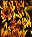
Porphyra Algae
Members of the Porphyra genus of algae are commonly consumed by humans. In nature, these red algae are typically found in temperate intertidal zones, but most Porphyra marketed today is cultivated at large sea farms in China, Japan, and South Korea.

The discovery that Porphyra exhibits a trimorphic life cycle in the late 1940s contributed greatly to the success of commercial operations, which had been up to that time under poor control since no one understood exactly how the algae multiplied. Indeed, for many years it had been assumed that the filamentous stage of the Porphyra life cycle was a different species that was named Conchocelis rosea. Now it is known, however, that filamentous morphs of Porphyra are formed following a carposporophyte stage exhibited by the genus, and that they develop into the familiar edible thallus form subsequent to this stage's maturation in the shells of marine animals, which leads to the release of conchospores.
Porphyra algae may be prepared in a variety of ways before they are eaten. One of the most common practices is to shred the blades and press them into sheets to dry. In this form, Porphyra is often known as nori, a food item that can be used to make sushi, soups, fried snacks, and other dishes especially popular in Asia. In the British Isles, Porphyra is typically referred to as laver or sloke and is eaten as a fresh vegetable, marinated and grilled on toast, as a jelly, or mixed with oatmeal and fried to make laver bread. Porphyra contains a variety of nutrients in significant amounts and is high in protein, but has yet to become widespread as a human food in North America, where it is sometimes alternatively utilized to feed saltwater fish by aquarium enthusiasts.
Contributing Authors
Nathan S. Claxton, Shannon H. Neaves, and Michael W. Davidson - National High Magnetic Field Laboratory, 1800 East Paul Dirac Dr., The Florida State University, Tallahassee, Florida, 32310.