 |
 |
 |
|
||||||||||||||||||||||||
 | ||||||||||||||||||||||||
 | ||||||||||||||||||||||||
 | ||||||||||||||||||||||||
Confocal Microscopy Image Gallery
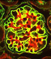
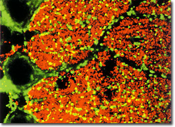
Rat Cerebellum Purkinje Cells
Purkinje cells in the rat cerebellum were stained with a dual fluorophore strategy to produce the beautiful confocal image presented below. The green fluorescent probe fluorescein isothiocyanate (FITC) was used to label vesicular gamma-aminobutyric acid transporter VGAT), while red Cy3 labeled the vesicular glutamate transporter (VGLUT1). The image was provided by Masahiko Watanabe from the Department of Anatomy at the Hokkaido University School of Medicine, in Japan.
Located posterior to the pons and medulla oblongata, the cerebllum, which means little brain in Latin, is a trilobed structure near the base of the brain. Its primary function is to regulate and control complex voluntary muscular movements, such as running or jumping. To do this, the cerebellum must also be able to smoothly coordinate information from various parts of the body, including the eyes, ears, limbs, and the cerebrum. The surface of the cerebellum is grooved and is composed of gray matter, while the structures interior consists of white matter. Within these layers are five different kinds of nerve cells, but of these only the branching Purkinje cells extend axons outside of the cerebellum.
Confocal microscopy is ideally suited for the study of Purkinje cells and other nerve tissue components due to its ability to facilitate the examination of thick specimens, such as brain slices. For example, by utilizing this technique with a variety of fluorescent dyes researchers have been able to successfully investigate calcium signaling and synaptic transmission in cerebellar slices, to image GABA-mediated changes in intracellular chloride within individual neurons, and to observe the morphological changes in Purkinje cells treated with thyroid hormones. Similar scientific findings have also been facilitated by the optical sectioning aspect of confocal microscopy, which can be utilized to create extremely detailed three-dimensional images of neural anatomy.