 |
 |
 |
|
||||||||||||||||||||||||
 | ||||||||||||||||||||||||
 | ||||||||||||||||||||||||
 | ||||||||||||||||||||||||
Confocal Microscopy Image Gallery
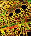
Zinnia elegans Mesophyll Cells
Isolated mesophyll cells from the flowering plant Zinnia elegans were multiply stained and reconstructed using volume rendering techniques to produce the image illustrated below. The image was provided by Keisuke Obara and Hiroo Fukuda from the Department of Biological Sciences in the Graduate School of Science at the University of Tokyo in Japan.

Predominantly native to North America, more than 20 species of herbs and shrubs are categorized in the genus Zinnia. Perennial where they are indigenous, the plants are characterized by lance-like or oval-shaped leaves that are rough in texture and which are arranged in opposition to one another along their relatively rigid, hairy stems. The Mexican species, Zinnia elegans, is the origin of the many showy, flowering varieties of the plant that are commonly found in gardens and flowerbeds. Easily cultivated, zinnias occur in an array of colors that range from pure white to bright red, yellow, and purple.
Confocal microscopy is an excellent method for examination of the fine details of plants, such as the irregularly shaped mesophyll cells that appear in the leaves of Zinnnia elegans and other angiosperms. Optical sectioning can facilitate the observation of living specimens, reconstruction enabling the creation of representative three-dimensional images that are anatomically correct. The images produced with this technique are especially refined because confocal laser scanning microscopes permit the exclusion of light that is out of focus. Also, fluorescent probes may be used to identify with tremendous accuracy the location and appearance of cellular components, such as mitochondria, chloroplasts, and peroxisomes.