 |
 |
 |
|
||||||||||||||||||||||||
 | ||||||||||||||||||||||||
 | ||||||||||||||||||||||||
 | ||||||||||||||||||||||||
Scanning Techniques

The Olympus FluoViewTM confocal microscopes provide innovative scanning techniques for improved performance.
The efficient Olympus multiple confocal scanning modes include point, line, free line and rectangle scanning, which make the FV300 and FV500 especially suitable for many time-lapse applications. In laser scanning confocal microscopy, the image of an extended specimen is generated by scanning the focused beam across a defined area in a raster pattern controlled by two high-speed oscillating mirrors driven by galvanometer motors. One of the mirrors moves the beam from left to right along the x lateral axis, while the other translates the beam in the y direction. After each single scan along the x axis, the beam is rapidly transported back to the starting point and shifted along the y axis to begin a new scan in a process termed flyback. During the flyback operation, image information is not collected. In this manner, the area of interest on the specimen in a single focal plane is excited by laser illumination from the scanning unit.

Software control of the scanning mode in the FluoViewTM confocal microscopes enables the operator to perform a wide variety of experiments. For example, in the frame on the right, a brain slice stained for the Golgi apparatus (black/white) and counterstained with Alexa Fluor 594 (red) for glial fibrillary acid protein (GFAP) demonstrates line (yellow line), rectangular (yellow box), and free line (blue) scanning mechanisms. Superimposed on the image is a selection palette from the FluoViewTM software that contains scanning mode controls. The image was provided by Drs. Matt Blurton-Jones and Mark Tuszynski from the University of California in San Diego.
Scanning Modes
- Line Scanning - In this technique, a single line may be scanned while oriented at any angle in the x-y plane. This fast scanning option permits accurate quantitative analysis of physiological events such as calcium waves or sparks.
- Rectangle Scanning - A scanned rectangular area can be rotated in the x-y plane, enabling efficient sampling of the specimen.
- Point Scanning - The ultimate in fast scanning, the point scan enables accurate quantitative analysis of intensity changes during rapid physiological events.
- Free Line Scanning - Intensity changes may be measured over a chosen time period along the length of a freely drawn line, such as the trace of an axon or along a cellular junction.

Sequential Scanning to Prevent Crosstalk

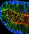
Sequential scanning is a technique introduced to confocal microscopy in order to minimize the crosstalk between channels, which often occurs in specimens that are multiply stained with two or more fluorophores having overlapping emission spectra. Individual spectra are collected from each dye using the FluoViewTM AOTF function and subsequently added together in the software. For example, the human colon crypt on the right was multiply stained with TO-PRO-3 (nuclei (a); blue), Alexa Fluor 488 phalloidin (actin (b); green), and Alexa Fluor 569 (APC gene product (c); red). Each fluorophore channel was collected separately and the images added together in the software to form a multi-spectral composite (d) that vividly displays all three colors with a minimum of crosstalk. The images were provided by Drs. Christine Anderson and Ray White of the Huntsman Cancer Institute at the University of Utah.