 |
 |
 |
|
||||||||||||||||||||||||
 | ||||||||||||||||||||||||
 | ||||||||||||||||||||||||
 | ||||||||||||||||||||||||
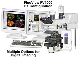
The Olympus FluoViewTM FV1000 is a next-generation imaging system designed for high-resolution, confocal observation of both fixed and living cells. The FV1000 offers advances in confocal system performance while providing the speed and sensitivity required for live cell imaging with minimal risk of damage to living specimens. In addition, the FV1000 offers a revolutionary synchronized laser scanning system called the SIM Scanner. While one laser stimulates, the second laser simultaneously provides high-resolution imaging. This coordination of laser stimulation and imaging makes the FV1000 an ideal choice for FRAP, FLIP and photoactivation.
The newly developed FV1000 scanning units offer a high-sensitivity detection system coupled to high-speed scanning and laser feedback to enable the capture of high-quality images of living organisms undergoing rapid change. Images may be scanned in any pixel array size up to 2048 x 2048, with each channel digitized to 4096 gray levels (12-bit resolution), permitting observation of fine image detail. The SIM scanner unit employs a new spectral imaging technology using diffraction gratings to provide distinct wavelength separation with precise resolution.

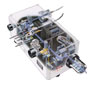
Included in the compactly-designed scanning units (see Figure 1) are a fiber optic coupler for multiple incoming laser light sources, a beam collimator, laser adjustment neutral density filter turret, dichromatic mirrors, polarizer, x-y galvanometer-driven scan mirrors, pinhole aperture, photomultipliers, beamsplitters, fluorescence filters, diffraction gratings, slits, and a collector lens.
SIM Scanner System Synchronizes Excitation and Imaging
In addition to the scanning laser for observation, the main FV1000 scanning unit incorporates a compact scanner for laser light excitation. Because the exchange between imaging and excitation involves no time lag, reactions such as fluorescence acquisition immediately following stimulation are not overlooked. This capability is applicable to many investigations, including recovery after photobleaching (FRAP), loss in photobleaching (FLIP), photoactivation, and uncaging.

Tornado scanning, a unique feature of the FV1000, increases photobleaching efficiency. Conventional reciprocating scanning systems tend to be slow, and there are cases when photobleaching is inadequate. However, with tornado scanning (which requires a SIM scanner to be loaded), there is no waste scanning, and photobleaching is much more efficient. The SIM scanning unit offers two galvanometer mirror scanners, a pupil projection lens, built-in laser shutter, and ports for incident illumination via a fiber connector for visible and ultraviolet lasers.
The highly sensitive detection system featured by the FV1000 minimizes cell damage. Dichromatic mirrors and barrier filters are made with ion deposition techniques for increased sensitivity and full wavelength coverage. These filters feature a multi-layer coating to provide sharp cut-on and cut-off characteristics, which cannot be achieved using conventional vacuum deposition methods. Another key advantage is the total coverage of all fluorescence wavelength ranges by the FV1000 filter turrets, including those exhibited by fluorescent proteins.

The FV1000 four-channel scanning unit features a unique multi-spectral detection system that is sensitive, fast, and accurate. Using an independent photomultiplier for each channel, the optical system of the scanning unit also contains diffraction gratings and slits for two of the detectors for spectroscopy. The spectral scanning system delivers outstanding performance without deviation in wavelength, enabling high-precision, high-resolution, and high-speed spectroscopy (acquisition of lambda stacks) in observations ranging in timescale between milliseconds and hours.
In order to separate two similar fluorescence wavelengths with the spectrometer FV1000 scanning unit, two modes are provided: Normal mode, which requests the input of spectral profile data; and Blind mode, which calculates the separation automatically based on information derived from the specimen image without any additional spectral data. Accurate separations can, therefore, be performed easilyeven for situations, such as autofluorescence, for which spectral profiles are unavailable. The FV1000 features 2-nanometer wavelength resolution, making it possible to clearly separate crosstalk produced by two fluorochromes with similar or overlapping spectra. In addition, images of specimens with fluorescence emission profiles similar to those of excitation can be acquired.

The FV1000 scanning units offer high-speed and precision detection when conducting time-lapse experiments. The units are capable of acquiring data at 16 frames per second with extremely stable galvanometer mirrors. A feedback mechanism, which monitors laser light in the scanner, feeds data back to the laser combiner's acousto-optic tunable filter (AOTF) to keep excitation light intensity constant during time-lapse experiments. A hybrid photon counting mode enables fluorescence intensity to be accurately measured. The Time Series Controller software is used to control the precision of time-lapse experiments in microseconds, which also enables very accurate control of synchronization with external devices. Low signal-to-noise detection for these investigations is afforded by a pupil projection lens, photomultipliers with high sensitivity, and a low-noise analog processing circuit, all integrated into the optical system.

The wide range of choices in FV1000 scanning units offers greater system diversity to allow easy expandability and operability. In most cases, the scanning unit with its integrated fluorescence illuminator is positioned at the rear of the microscope main body, leaving free space on both sides to be utilized as required. In addition, the side port can be coupled to the scanning head in an IX81 combination, as well as the video port of the intermediate magnification unit in a BX61WI combination.