Classification and Applications of Fluorescent Probes
Fluorescent probes used in confocal microscopy are classified based on spectral properties (excitation/emission), chemical structure, target specificity, and functional application. These probes enable high-resolution imaging of cellular structures, molecular interactions, and dynamic physiological processes in both fixed and live specimens.
1.Biomolecule-Specific Probes
(Proteins, nucleic acids, lipids, carbohydrates, toxins)
Receptor Ligands (Agonists/Antagonists)
TRITC–α-bungarotoxin
Specifically binds acetylcholine receptors at neuromuscular junctions to visualize receptor distribution.
Antibodies (Immunofluorescence)
FITC-labeled secondary antibodies (anti-IgG / IgM)
Enable indirect detection of primary antibodies for highly specific protein localization.
Biotin–Avidin Detection Systems
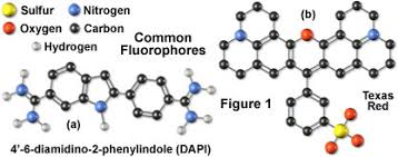
Texas Red–streptavidin (avidin)
Detects biotinylated probes (e.g., FISH probes, antibodies) via strong biotin–avidin binding.
Dextran-Conjugated Probes
Texas Red–dextran
High-molecular-weight polysaccharide used for cytoplasmic tracing, ion sensing, or pH measurements after microinjection.
Fluorescent Lipid Analogues
NBD–phosphocholine
Incorporated into membranes to study membrane dynamics, fusion, and fluidity (e.g., FRAP analysis).
Fluorescent Nucleotides
Cy3–dCTP
Used for DNA labeling in fluorescence in situ hybridization (FISH) via nick translation.
Alternative tags: biotin- or digoxigenin-labeled nucleotides.
Cytoskeletal Probes
Rhodamine–phalloidin
Specifically binds F-actin for visualization of actin filaments and stress fibers.
2. Membrane Imaging Probes
Voltage-Sensitive Dyes
WW 781, RH-155, Di-8-ANEPPS
Rapid-response dyes that change fluorescence properties according to membrane potential.
Organelle-Specific Membrane Probes
DiOC6(3) – Endoplasmic reticulum
NBD–ceramide – Golgi apparatus
Rhodamine 123 – Mitochondria
Lipophilic Tracers
DiI, DiO, DiA
Track membrane incorporation and neuronal projections.
3. Cytoplasmic and Functional Probes
Ion-Sensitive Indicators
Fura-2, Indo-1, Fluo-3 – Calcium imaging
BCDCF, SNARF-1 – pH measurement
Often delivered as membrane-permeable AM esters for live-cell studies.
Tracer Dyes
Sulforhodamine 101, Lucifer Yellow
Used for microinjection-based cell tracing and neuronal connectivity studies.
Live-Cell Viability Probes
Calcein AM, CFDA
Converted by intracellular esterases into fluorescent, membrane-impermeable forms, enabling viability assessment.
4. Nuclear and Genetic Probes
DNA/RNA Stains
DAPI
Propidium iodide (PI)
Hoechst 33342
YOYO-1
Bind nucleic acids for nuclear morphology, chromosome analysis, and DNA quantification.
Fluorescent Proteins
GFP (Green Fluorescent Protein) and variants
Genetically encoded reporters for live-cell imaging of protein localization, gene expression, and intracellular dynamics.
5️. Functional and Enzymatic Probes
Enzyme-Activated Dyes
Dihydrorhodamine 123
Becomes fluorescent upon enzymatic oxidation; used to monitor intracellular enzymatic activity and oxidative stress.
6️. Fluorescent Particles and Calibration Tools
Fluorescent Latex Beads
FluoSpheres
Used for axonal transport studies, phagocytosis assays, blood flow measurements, and system calibration (resolution standards).
7. Key Applications in Confocal Microscopy
Confocal fluorescence imaging enables:
High-resolution subcellular localization
3D reconstruction of tissues
Live-cell dynamic imaging
Ion flux and pH monitoring
Membrane trafficking and fusion studies
Cytoskeletal architecture analysis
Gene expression and chromosomal mapping
Signal transduction and enzymatic activity measurement
Quantitative fluorescence analysis