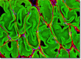
Rat Diaphragm Smooth Muscle Tissue
Presented above is a laser scanning confocal micrograph illustrating the extensive filamentous actin network within smooth muscle tissue from an 8-µm cryosection of rat diaphragm.
The specimen was labeled with a dual-fluorophore cocktail consisting of Alexa Fluor 488–conjugated phalloidin to visualize F-actin and Texas Red-X–conjugated wheat germ agglutinin (WGA) to label lectin-binding glycoconjugates. Cell nuclei were counterstained with Hoechst 33342.
Images were acquired in grayscale using an Olympus FluoView FV1000 confocal system mounted on a BX-81 inverted microscope, with excitation provided by an argon-ion laser (488 nm), a violet diode laser (405 nm), and a green helium–neon laser (543 nm). During post-acquisition processing, individual fluorescence channels were pseudocolored using RGB assignments corresponding to the emission spectra of each fluorophore and merged to generate the final composite image.