Immunofluorescence Imaging of a Rat Brain Section
The lateral ventricles are the largest components of the brain’s ventricular system, a network of interconnected cavities that circulate cerebrospinal fluid (CSF). CSF provides mechanical protection, nutrient transport, and metabolic waste removal. Each lateral ventricle has a characteristic C-shape with three major extensions: the anterior horn (frontal lobe), posterior horn (occipital lobe), and inferior horn (temporal lobe). The junction of these regions is termed the atrium.
CSF is primarily produced by the choroid plexus located within the lateral ventricles, with additional contributions from the third and fourth ventricles. Fluid exits the system through apertures in the fourth ventricle and is absorbed into the subarachnoid space. Obstruction of CSF flow can lead to hydrocephalus, characterized by ventricular enlargement and increased intracranial pressure. In infants, this may cause cranial expansion due to unfused sutures, whereas in older individuals symptoms include headache, nausea, visual disturbances, gait instability, and cognitive impairment. Untreated hydrocephalus is life-threatening but can be managed surgically via shunt placement or ventriculostomy.
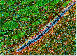
The lateral ventricle is visible in the coronal thin section of rat brain shown above. The specimen was immunolabeled for:
Neuronal structures: heavy chain neurofilament (NF-H) using chicken anti-NF-H primary antibodies, detected with goat anti-chicken secondary antibodies conjugated to Alexa Fluor 488 (green emission).
Astroglial cells and neural stem cells: glial fibrillary acidic protein (GFAP) using rabbit anti-GFAP primary antibodies, detected with goat anti-rabbit secondary antibodies conjugated to Alexa Fluor 568 (red emission).
Nuclear DNA: counterstained with DRAQ5 (pseudocolored cyan).
Images were acquired using a 20× objective (zoom factor 1.3) with sequential laser scanning at 488 nm (argon-ion), 543 nm (green helium–neon), and 633 nm (red helium–neon). Individual fluorescence channels were recorded in grayscale and subsequently pseudocolored according to their respective emission spectra to produce the final composite image.