Mitochondria and Nuclear DNA in Bovine Pulmonary Artery Endothelial Cells
Visualization of Mitochondria and Nuclear DNA in Bovine Pulmonary Artery Endothelial Cells
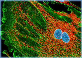
Bovine pulmonary artery endothelial (BPAE) cells were cultured and subjected to dual fluorescent labeling to simultaneously visualize the mitochondrial network and nuclear DNA. Mitochondria were stained with MitoTracker Red CMXRos, a rosamine-based fluorophore that selectively accumulates in active mitochondria due to their membrane potential. Nuclear DNA was counterstained with DRAQ5, a far-red, cell-permeable dye that binds stoichiometrically to chromatin, providing a clear nuclear signal without interfering with other fluorophores.
The dual-labeling approach enables detailed assessment of cellular architecture. Mitochondria appeared as discrete, highly interconnected filaments distributed throughout the cytoplasm, reflecting the dynamic and reticulated nature of the organelle network. In contrast, DRAQ5 produced a sharply defined nuclear signal, highlighting chromatin organization and allowing easy distinction of nuclei from the surrounding cytoplasm.
By combining these two fluorescent markers, researchers can evaluate mitochondrial morphology, distribution, and integrity in relation to nuclear organization. This is particularly valuable in studies of cellular metabolism, mitochondrial dynamics, and organelle crosstalk in vascular endothelial cells. Imaging of the labeled cells provides high contrast between cytoplasmic and nuclear structures, enabling quantitative or qualitative analyses of organelle interactions, spatial organization, and potential changes under experimental conditions.