Combined Fluorescence and Contrast Microscopy to Reduce Photobleaching
To minimize photobleaching, fluorescence imaging is often combined with non-destructive transmitted-light techniques such as differential interference contrast (DIC), phase contrast, Hoffman modulation contrast (HMC), and transmitted darkfield illumination.
The strategy is straightforward:
Locate and focus on the region of interest using a transmitted-light contrast method (which does not excite fluorophores).
Without moving the specimen, switch to fluorescence mode for targeted imaging.
This approach significantly reduces unnecessary fluorochrome exposure while preserving structural context.
Practical Example of Combined Imaging
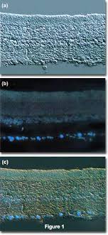
In a typical experiment, a thin section of rat retina optic ganglion tissue can first be visualized using DIC to reveal fine structural detail. The same field is then imaged under fluorescence illumination using a mercury vapor lamp and an appropriate filter cube, with cells stained by fast blue, a diazonium dye that selectively labels phospholipids in the myelin sheath.
When both imaging modes are superimposed, fluorescence highlights the labeled lipid structures while DIC provides detailed morphological background. This dual-modality imaging is often achieved using specialized fluorite objectives designed to support both fluorescence and DIC simultaneously.
Optical Configuration for Simultaneous DIC and Fluorescence
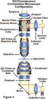
A microscope configured for combined imaging typically includes:
🔹 Differential Interference Contrast (DIC) Pathway
Transmitted light from a tungsten–halogen lamp
Field diaphragm and condenser optics
Wollaston prism in the condenser front focal plane
Second Wollaston prism near the objective rear focal plane
Interference at the intermediate image plane to produce high-contrast structural detail
🔹 Fluorescence Pathway
Ultraviolet excitation from a mercury burner
Exciter filter to select excitation wavelength
Dichroic mirror to reflect excitation light onto the specimen
Emission (secondary fluorescence) collected by the objective
Barrier (emission) filter to isolate fluorescence signal
Detection via eyepieces or phototube
Both imaging modes can be used independently or simultaneously, allowing structural and molecular information to be visualized within the same specimen and focal plane.
Advantages of Combined Imaging
Reduced photobleaching
Improved localization accuracy
Enhanced structural context
Efficient targeting of regions of interest
Ideal for live-cell and sensitive specimens