 |
 |
 |
|
||||||||||||||||||||||||
 | ||||||||||||||||||||||||
 | ||||||||||||||||||||||||
 | ||||||||||||||||||||||||
Confocal Microscopy Image Gallery
Indian Muntjac Deer Skin Fibroblast Cells
A fibroblast cell line established from a skin biopsy of an adult male, the Indian Muntjac deer epidermis line is commonly used in laboratories around the world, especially for chromosome studies. Members of the family Cervidae, Muntjacs are barking deer that emit their characteristic sound when they feel threatened or alarmed.

The normal (non-transformed) Indian Muntjac cell line is susceptible to the herpes simplex virus, vaccinia virus, and vesicular stomatitis virus (Indiana strain), but is resistant to poliovirus 1. Recent tests have demonstrated that the cells produce both detectable bovine viral diarrhea virus (BVDV) antigens and infectious BVDV virions. Muntjac cells are negative for reverse transcriptase, indicating the lack of integral retrovirus genomes.
Cell lines derived from the Indian Muntjac have been of significant scientific interest primarily because the animal possesses the fewest number of diploid chromosomes of all mammals, with only six chromosomes in the female and seven in the male. Such a small number of chromosomes makes Indian Muntjac cells an ideal candidate for mitosis research. Moreover, in recent years, Indian Muntjac cells have gained a reputation for their usefulness as a model to study telomere biology. Telomeres, the regions of DNA that occur at the end of chromosomes, are at the center of many modern studies involving the aging of organisms and cell senescence. It is generally believed that the shortening of telomeres, which occurs during the division of most cells, is responsible for age-related cellular malfunction and death. Thus, telomeres are also of interest in regard to cancer, since tumor cells are often able to proliferate unhindered by the passage of time due to various mechanisms that avoid the shortening of chromosomal telomeres.
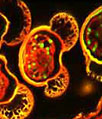
The Indian Muntjac deer skin fibroblast cells presented in the digital image above were resident in an adherent culture stained for F-actin with Alexa Fluor 546 conjugated to phalloidin, and for DNA with the red-absorbing dye TO-PRO-3 (pseudocolored blue). In addition, the culture was immunofluorescently labeled with Alexa Fluor 488 conjugated to antibodies that target alpha-tubulin, a major component of the microtubule network. Images were recorded on a FluoView FV1000 with a 60x oil immersion objective using a zoom factor of 2.0 and sequential scanning with the 488-nanometer spectral line of an argon-ion laser, the 543-nanometer line from a green helium-neon laser, and the 633-nanometer line of a red helium-neon laser. During the processing stage, Alexa Fluor image channels were pseudocolored with RGB values corresponding to each of the fluorophore emission spectral profiles.
Additional Confocal Images of Indian Muntjac Cells
Imaging the Mitochondria Network, Filamentous Actin, and Nuclei in Indian Muntjac Deer Skin Fibroblast Cells - A triple fluorophore combination of MitoTracker Deep Red 633, BODIPY FL conjugated to phalloidin, and SYTOX Orange was used to label an adherent log phase culture of Indian Muntjac cells for mitochondria, the filamentous actin network, and nuclear DNA, respectively. The cells were first treated with MitoTracker Deep Red 633 in growth medium for one hour, washed and fixed with paraformaldehyde (prepared in growth medium), permeabilized, and blocked with bovine serum albumen. The cells were subsequently labeled with the conjugated phalloidin and counterstained with the SYTOX reagent.
The Mitotic Apparatus in Dividing Indian Muntjac Deer Skin Cells - The first stages of mitosis are revealed in the confocal image of Indian Muntjac deer skin cells presented in this section, which were stained with Alexa Fluor 568 (tubulin) and TO-PRO-3 (DNA). The cells were fixed with 3.7 percent paraformaldehyde, permeabilized with Triton X-100, blocked with 10 percent normal goat serum and treated with mouse anti-alpha-tubulin primary antibodies. Goat anti-mouse secondary antibodies conjugated to Alexa Fluor 568 were mixed with the DNA stain.
Indian Muntjac Cells with Alexa Fluor 488 and TO-PRO-3 - An adherent culture of Indian Muntjac deer skin fibroblast cells was fixed, permeabilized, blocked, and then treated with mouse primary antibodies that target alpha-tubulin, a major component of the microtubule network. The primaries were labeled with goat anti-mouse secondary antibodies conjugated to Alexa Fluor 488 in a solution containing TO-PRO-3 to simultaneously stain DNA.
MitoTracker, Actin, and DNA Probes with Indian Muntjac Deer Skin Fibroblast Cells - A triple fluorophore combination of MitoTracker Orange CMTMRos, BODIPY FL conjugated to phalloidin, and TO-PRO-3 was used to label an adherent log phase culture of Indian Muntjac cells for mitochondria, the filamentous actin network, and nuclear DNA, respectively. The cells were first treated with the MitoTracker probe in growth medium for one hour, washed and fixed with paraformaldehyde (prepared in growth medium), permeabilized, and blocked with bovine serum albumen. The cells were subsequently labeled with the conjugated phalloidin and counterstained with the cyanine monomer (TO-PRO-3) reagent.
The Metaphase Spindle in Dividing Indian Muntjac Cells - The metaphase spindle apparatus is revealed in this confocal image of Indian Muntjac deer skin cells stained with Alexa Fluor 568 for microtubules and TO-PRO-3 for DNA. The cells were fixed with 3.7 percent paraformaldehyde, permeabilized with Triton X-100, blocked with 10 percent normal goat serum and treated with mouse anti-alpha-tubulin primary antibodies. Goat anti-mouse secondary antibodies conjugated to Alexa Fluor 568 were mixed with the DNA stain.
Indian Muntjac Fibroblasts with MitoTracker Red CMXRos, Alexa Fluor 488, and DRAQ5 - The cytoskeletal F-actin and mitochondrial networks were targeted in the culture of Indian Muntjac cells presented in this section with Alexa Fluor 488 (pseudocolored blue) conjugated to phalloidin (a mushroom toxin) and MitoTracker Red CMXRos, respectively. Nuclei in the fibroblasts were counterstained with the far-red fluorescent DNA probe DRAQ5 (pseudocolored yellow).
Distribution of F-Actin and Mitochondria in Muntjac Cell Cultures - The proximity between the mitochondrial network and the filamentous actin cytoskeleton was visualized by treating a culture of Indian Muntjac deer skin fibroblasts with MitoTracker Red CMXRos and Alexa Fluor 488 conjugated to phalloidin. The specimen was also probed with DRAQ5, a DNA-interactive reagent that exhibits preferential intercalation at AT base pairs.
Targeting the Mitochondria Network and Nuclear DNA in Indian Muntjac Deer Skin Fibroblasts - The monolayer culture of Indian Muntjac fibroblast cells shown in this section was treated with MitoTracker Red CMXRos in medium containing 15 percent Cosmic calf serum, fixed with the same medium containing 3.7 percent paraformaldehyde, permeabilized with 0.2 percent Triton X-100, and then counterstained with DRAQ5 (pseudocolored cyan), targeting DNA in the cell nuclei.
Muntjac Skin Cells Labeled for Tubulin with Immunofluorescence - In order to visualize the microtubules present in a culture of Indian Muntjac fibroblasts, the cells were immunofluorescently labeled with anti-tubulin mouse monoclonal primary antibodies followed by goat anti-mouse Fab fragments conjugated to Alexa Fluor 488. In addition, the cells were labeled for mitochondria with MitoTracker Red CMXRos, a derivative of X-rosamine, and DNA in the nucleus with the far-red fluorescent probe DRAQ5.
Visualizing the Nuclear Histones and Cytoplasmic Peroxisomes in Indian Muntjac Cell Cultures - A log phase culture of Indian Muntjac cells was treated with a cocktail of mouse anti-histones (pan) and rabbit anti-PMP 70 (peroxisomal membrane protein) primary antibodies, followed by goat anti-mouse and anti-rabbit secondary antibodies conjugated to Alexa Fluor 647 and Alexa Fluor 488, respectively, to target the nuclear histone proteins and peroxisomes. In addition, the F-actin network was labeled with Alexa Fluor 568 conjugated to phalloidin, a phallotoxin isolated from the death cap mushroom.
Indian Muntjac Deer Skin Cells Triple Labeled for Mitochondria, Filamentous Actin, and DNA - A triple fluorophore combination of MitoTracker Red CMXRos, Alexa Fluor 488 conjugated to phalloidin, and DRAQ5 was used to label the adherent culture of Indian Muntjac cells presented in this section for mitochondria, the F-actin network, and nuclear DNA, respectively. The cells were first treated with the MitoTracker probe in growth medium for one hour, washed and fixed with paraformaldehyde (prepared in growth medium), permeabilized, and blocked with bovine serum albumen. The cells were subsequently labeled with the conjugated phalloidin and counterstained with the anthraquinone (DRAQ5) reagent.
Peroxisome and Histone Protein Distribution in Muntjac Fibroblasts - In a double immunofluorescence labeling experiment, a culture of Indian Muntjac fibroblasts was treated with a cocktail of mouse anti-histones (pan) and rabbit anti-PMP 70 (peroxisomal membrane protein) primary antibodies, followed by goat anti-mouse and anti-rabbit secondary antibodies conjugated to Alexa Fluor 568 and Alexa Fluor 488, respectively, to target the nuclear histone proteins and peroxisomes. The actin cytoskeleton was visualized with Alexa Fluor 633 conjugated to phalloidin.
Indian Muntjac Cells with MitoTracker Red CMXRos, Alexa Fluor 488, and DRAQ 5 - The adherent monolayer Indian Muntjac cell culture shown in this confocal image was labeled for the cytoskeletal filamentous actin and intracellular mitochondrial networks with Alexa Fluor 488 conjugated to phalloidin and MitoTracker Red CMXRos, respectively. Nuclei present in the fibroblasts were counterstained with the far-red fluorescent DNA-probe, DRAQ 5.
Contributing Authors
Nathan S. Claxton, Shannon H. Neaves, and Michael W. Davidson - National High Magnetic Field Laboratory, 1800 East Paul Dirac Dr., The Florida State University, Tallahassee, Florida, 32310.